Neurofibromatosis type 1
Management of neurofibromatosis type 1Lena (9 years old)
Lena is a 9-year-old girl with neurofibromatosis type 1 (NF1), under follow-up by a local paediatric neurologist. Over the past year, she has developed a progressive swelling below the left mandibular angle, which she increasingly perceives as a bothersome lump. Following MRI of the head and neck, she was first referred to a paediatric NF1 centre, followed by referral to another NF1 centre with expertise in peripheral nerve surgery.
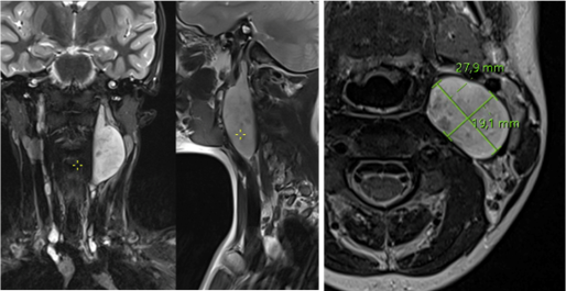
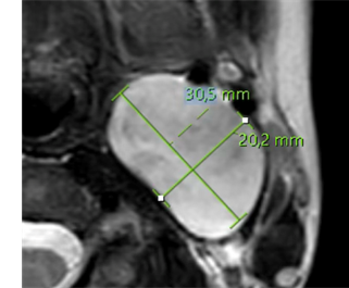
A follow-up MRI 6 months later demonstrated tumour growth of 2-3 mm in all dimensions.
Assessment summary:
- Medical history:
- NF1 diagnosis in early childhood based on increasing café-au-lait macules and axillary freckling, with subsequent genetic confirmation of a pathogenic NF1 mutation
- Annual follow-up including paediatric clinical, neurological and dermatological assessment, ophthalmological assessment of visual function, and abdominal ultrasound
- Symptoms: progressive swelling below the left mandibular angle, increasingly perceived as a foreign body sensation, with mild pain and discomfort on deep palpation
- Normal swallowing and unchanged normal voice
- Follow-up MRI: mild plexiform neurofibromatous transformation of a nerve within the neurovascular sheath of the left cervical region, with a large 3x2x3cm intraneural distinct nodular lesion above the carotid bifurcation, located between the trachea and oesophagus medially, the 2 carotid arteries anterolaterally and the severely compressed jugular vein posterolaterally
First MRI Second MRI, 6 months later
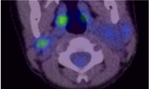
PET-CT, 1 month after second MRI